Closed sr320 closed 7 years ago
Final list:
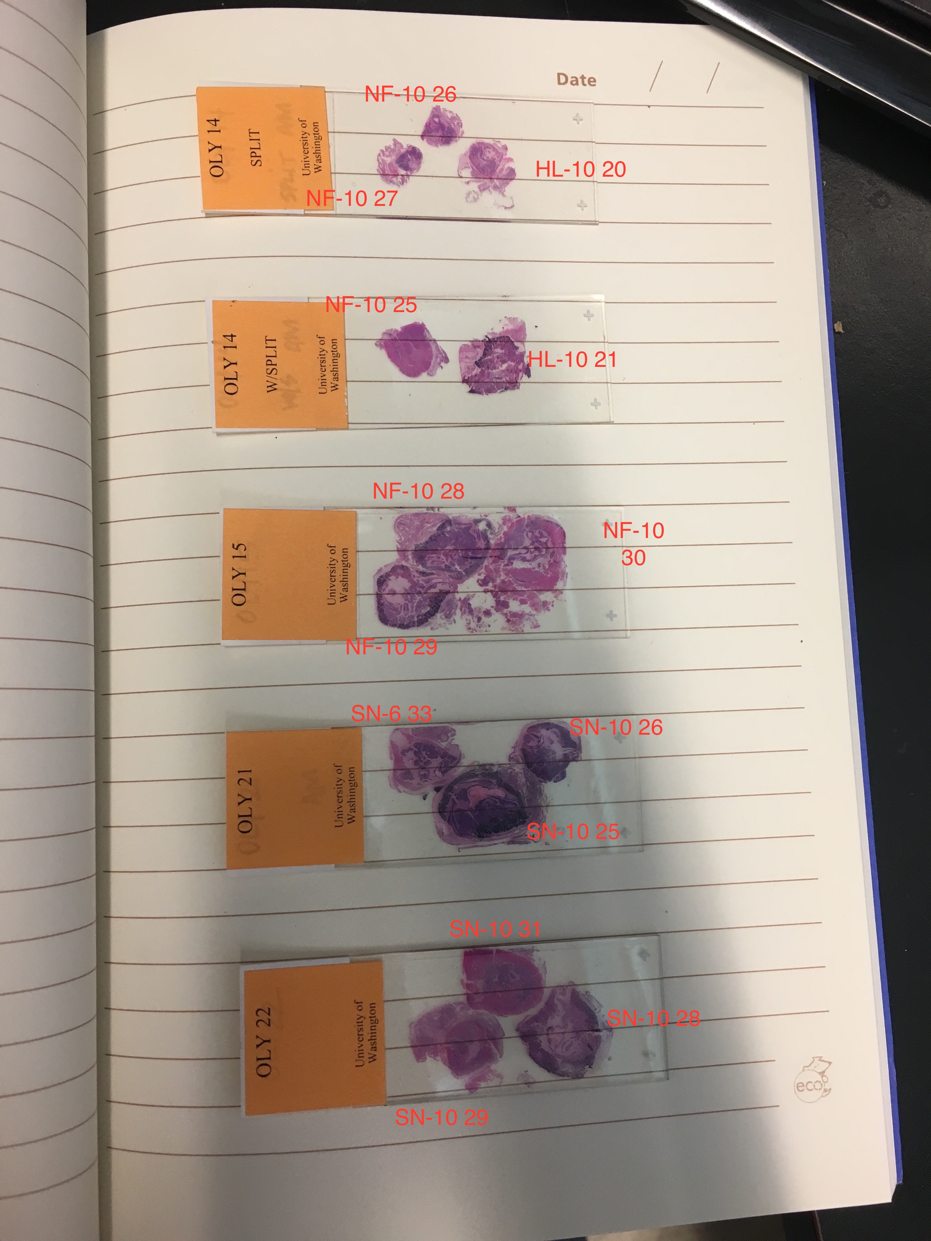
SN low pH
SN ambient pH
NF low pH
NF ambient pH
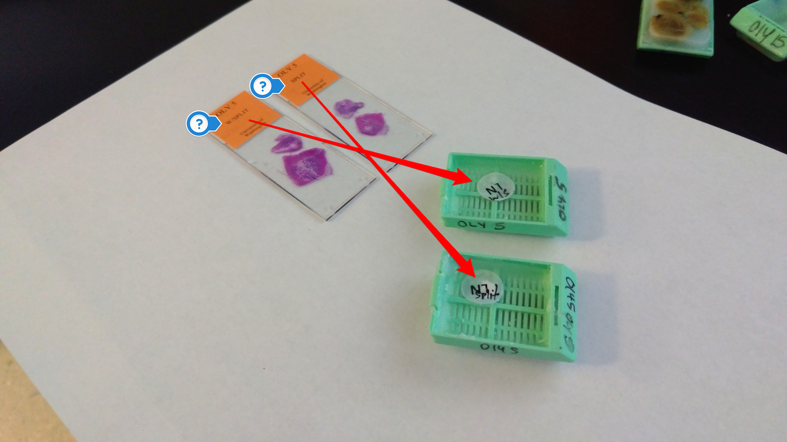
@kubu4, you'll probably want to work with Katie to make sure that you're extracting from the same sample as the one she imaged. The histology folks did not keep the samples in the same formation. Katie used my images of the prepared cassettes and my histology "map" to ID each sample.
Apparently, I can't tag Katherine in this repo.
Any idea how much RNA she needs from each sample?
@katiedavidson5 - I saw in your notebook that you indicated you uploaded your labeled images to "the google drive", but there's no link (and, I don't know what Google Drive you're referring to). Could you please drop a link here (and in your notebook)?
See https://genefish.wordpress.com/2017/06/28/katies-notebook-imaging-627/ On Wed, Jul 5, 2017 at 7:23 AM kubu4 notifications@github.com wrote:
@katiedavidson5 https://github.com/katiedavidson5 - I saw in your notebook that you indicated you uploaded your labeled images to "the google drive", but there's no link (and, I don't know what Google Drive you're referring to). Could you please drop a link here (and in your notebook)?
— You are receiving this because you authored the thread. Reply to this email directly, view it on GitHub https://github.com/sr320/LabDocs/issues/648#issuecomment-313118155, or mute the thread https://github.com/notifications/unsubscribe-auth/AEPHtyluv0il7V_Y--x-Lsf6MUlLxYFlks5sK5w2gaJpZM4OLFWV .
Thanks! Didn't go back that far in time.
OK, @laurahspencer and @katiedavidson5, I'm definitely going to need some help with these. Here's one example. Oly 5 block looks like this:
But, the original cassette looks like this:
@katiedavidson5 has mapped most of these back and ID -
She has certainly documented which ones she cannot ID https://genefish.wordpress.com/author/katiedavidson5/
But I presume the list (picked by @laurahspencer) could be ID'd by the fact she provided info based on @katiedavidson5 pics
Agreed. I am using @laurahspencer's list. The example posted above should contain NF-10-22 and NF-10-23 (which are listed as Samples 9 & 8 in @laurahspencer's post above), as well as NF-10-20 and NF-10-21.
I'm also not seeing any images of the histology blocks being mapped to the original cassette layouts. This is important, as it seems that the histo blocks are not in the same layout as they were in the cassette (looks like most of them have been flipped 180 degrees on the vertical axis; i.e. "mirrored" layout).
I think all of @katiedavidson5's images are only of slides - not mapping blocks to cassettes.
OK, I think I'm getting things figured out. Some of these cassettes were split into two different histo blocks (I'm guessing by the histology facility). I figured this out when looking at the slide labels. See images below of the Oly 5 block(s) for an example:
@kubu4 sorry for the delay; did you see these?:
images of the prepared cassettes prior to sending to histology and my histology "map" of the prepared cassettes.
It's confusing, as the histology folks move these around. I applied blue ink on the top left tissue sample in each cassette (see images for location). Katie used my images to compare the share of each tissue in the cassettes to the slides.
@laurahspencer, yes, I have been looking at those. Did you see my post above, documenting some of the confusion/difficulty involved with getting this stuff figured out?
Katie compared the shape of each tissue in the blocks/slides to my imaged slides. @katiedavidson5, did you keep record of how you assigned the tissues their record numbers? If not, you'll have to do the same image comparison, for example, this is how I would assign NF-10 22 & 23:
For some back story (probably TMI)- I selected the samples based on their sex/maturation targeting a male, female, and hermaphrodite from each of the following groups: NF-10-low, NF-10-ambient, SN-10-low, & SN-10-ambient.
Thanks. I figured this out already (see my post above).
I was hoping that these had all been mapped out already, just like your example above.
@laurahspencer - Do you have an image of the Oly 14 cassette (I'm not seeing it in your notebook with the rest of the cassettes)? I don't need it to figure out the samples, but would be nice to have for my notebook.
@laurahspencer - Actually, I can't determine Oly 14 samples without the original cassette image. Sorry!
@laurahspencer - I've annotated the set of histology blocks that contain the samples in your list (except for blocks Oly 14, due to missing tissue cassette image). Please review my annotations and confirm (or disconfirm) they are correct:
Thanks!
FYI I found an image of block 14 (along with 12 and 13), and updated the images on my notebook post.
I started to go through the annotations, and it's super hard for me to confidently ID them from images; I'll need to do it in person on Friday when I'm in. Will that work for you? I do think there are a couple that you swapped, for example:
And in the "Oly 10" block the blue ink leads me to believe things need to be swapped. NOTE: I just checked the slide images that Katie took and it looks like she ID'd the pink tissue SN-10-21.
Friday is fine. All the slides and blocks are at my computer. I won't be in. Just provide me with annotated images of the blocks and I'll get the RNA isolated.
Will do!
Sorry for the late response! I will be in on Friday around 1pm and can help as well! I did not record the new locations of the oysters on all of the slides. I did for cassettes 24-31, and a few other confusing ones but that is it. Shouldn't be too hard for me to go through and label them as I imaged them though if there is any confusion!
Not that the histology place can help us with this particular set, but I've emailed them about the fact that a subset of the blocks have had the tissue re-arranged from how it was submitted to them. In fact, tissue rearrangement only occurred on a subset of blocks - all blocks with a "YL" sticker on them did not retain the tissue arrangement as it was submitted to them.
Okay here are the labeled slides as imaged! If they're positioned in the same orientation as the cassettes (like they are set up now on @kubu4 's desk) it'll be easy to tell which is which.
**far left oyster on OLY 6 slide in NF-10 24 (I left off the 24)
I double checked Katie's annotations and I concur with the above images. You should be all set for Monday - but please call me if you run into issues. I'll be @ the hatchery but can step away if needed. For example, if you run into an issue with one specimen and need me to quickly ID a replacement, I can do so.
Out of curiosity, have you extracted RNA from histology blocks before, and in doing so do you target only the tissue in question, for this situation gonad? I'm curious, since the slides show that some specimens have other tissue types amongst the gonad, but I did my best to select samples that had very well developed gonad, so hopefully it won't be too difficult if that's your aim.
Yes I've done this before. I will not be reviewing the slides prior to extractions. So, I will isolate from a random area within each designated tissue. If there's a specific region within each tissue that you'd like me to target, you'll need to annotate images of the blocks, with the desired region circled within each tissue.
Curious... did @laurahspencer indicate where gonad was in each sample or were samples random?
Samples were "random" (i.e. in the center of each designated tissue, in order to avoid accidental cross contamination from neighboring tissue).
Just saw this- I assume it's too late for me to indicate gonad locations?
On Mon, Jul 10, 2017 at 2:48 PM kubu4 notifications@github.com wrote:
Samples were "random" (i.e. in the center of each designated tissue, in order to avoid accidental cross contamination from neighboring tissue).
— You are receiving this because you were mentioned.
Reply to this email directly, view it on GitHub https://github.com/sr320/LabDocs/issues/648#issuecomment-314259308, or mute the thread https://github.com/notifications/unsubscribe-auth/AQdwfTESkqgYNdw7emxiMtzaejQMFUoRks5sMpv8gaJpZM4OLFWV .
Yep.
Shoot- I was in the weeds and just now came up for air. Is it feasible for me to see which area you sampled, aka is it obvious when looking at the histology block? I'll be in tomorrow and can check it out.
I took a look at the images - these tissue samples are of whole visceral mass, so on a couple of the samples you probably got more digestive gland than gonad; gonad is more prevalent on the perimeter of the samples. I will be in tomorrow and can take a closer look, and we can discuss.
@laurahspencer determine which ones are gonad (if any)
Mark embedded samples where gonad tissue can be obtained. Maybe circle with pencil?
Annotating the blocks digitally would be sufficient, however, if you were going to write directly on the blocks, I think the wax in the blocks is too soft for a pencil. Could you use thin tip marker on blocks?
@laurahspencer - was this done?
Grace is on it today!
On Wed, Jul 12, 2017 at 1:17 PM Steven Roberts notifications@github.com wrote:
@laurahspencer https://github.com/laurahspencer - was this done?
— You are receiving this because you were mentioned.
Reply to this email directly, view it on GitHub https://github.com/sr320/LabDocs/issues/648#issuecomment-314884083, or mute the thread https://github.com/notifications/unsubscribe-auth/AQdwfTYGJrvjhxxUv6zKR3E1duZwtiDFks5sNSmXgaJpZM4OLFWV .
Notebook entry link comparing the histology block extraction sites with the actual locations of gonad tissue : here
Samples were taken from the center, whereas gonad tissue is found around the edges. Gonad tissue locations (determined through use of microscope) are indicated by arrows.
More direct link: https://genefish.wordpress.com/author/graceac9/
@grace-ac i see that you have a couple comments on wrong Oly ID- did you see Katie's map posted on this thread 5 days ago?
@laurahspencer about to add final solution to your samples - will look at katie's map after!
fixed it!
For Katherine's libraries..
(Paxgene kit is here)
Samples are by scope in lab.
SN low pH male - SN-10-20 female - SN-10-16 hermaphrodite - SN-10-17
SN ambient pH
NF low pH
NF ambient pH - (missing image for NF-10-25)